The functions of the plasmolemma
1. Receptive function (interaction with hormones, mediators, and other chemical factors).
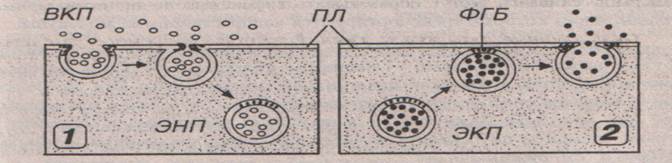
2. Transport function (transport of the substances into the cell is called endocytosis, from the cell- exocytosis.
- Endocytosis – the invaginated cell membrane fuses to form an endocytotic vesicle or endosome, which is a small, spherical membrane-bound body. The membrane and any material incorporated into such a vesicle can then be processed within the cell.
- Exocytosis is the reverse of endocytosis, and describes the fusion of a membrane-bound vesicle with the cell surface to discharge its contents into the extra-cellular space).
3. The formation of the intercellular contacts:
- Simple contact – membranes of two cells are on distance of 10 – 12 nm in such manner that the glycocalix one cell adjoins with glycocalix of another cell. The basic function is metabolism and information interchange between cells.

- Zonular occludentes – also known as tight junctions. Zonula occludens are located between adjacent plasma membranes most typically near the apices of epithelial cells. At the fusion sites occludins trans-membrane junctional proteins, lies. Occludin have more active role because these are the proteins that are probably responsible for the obliteration of the intercellular space by forming the tight junction. These junctions act as a barriers that prevent the movement of molecules into the intercellular spaces.

- Zonular adherents are belt-like junctions that assist adjoining cells to adhere to one another. This junction not only joins the cell membranes to each other but also links the cytoskeleton of the cells via the trans-membrane linker proteins. Apart from epithelial cells zonular adherents are also seen between smooth muscle cells, and between myocytes of cardiac muscle in the region of intercalated discs.

- Desmosomes. This is the most common type of tight junction between adjoining cells. At the side of a desmosome the plasma membrane (of each cell) is thickened because of the presence of dense layer of protein on its inner surface. The region of the gap is rich in a glycoprotein called desmoglein. The thickened areas of two membranes are held together by intermediate filaments of cytokeratin that appear to pass from one membrane to the other across the gap. Desmosomes are present where strong anchorage between cells is needed.

- Gap junctions, also called nexus or communicating junctions, are regions of intercellular communication. They are widespread in epithelial tissues, in cardiac muscle cells, smooth muscle cells and neurons. Gap junctions are built by six closely packed trans-membrane proteins connexins that assemble to from structures called connexons, aqueous pores through the plasma membrane extending into the intercellular space. The two connexons fuse, forming the functional communication channel. The hydrophilic channel permits the passage of ions, amino acids, small molecules and hormones.

- Synapse – type of contact between two nervous cells or between a nervous cell and a muscle. Through synapses pass nervous impulses.
Except for cells, multicellular organisms contain noncellular structures, which always are derivates of cells or products of their secretion. The symplastos, sincytium are postcellular structures.
The symplastos is a large formation with the big mass of cytoplasm and a plenty of nucleus (more ten). An example of symplastos are striated muscle fibers and external layer of placenta trophoblast.
The sincytium is a formation, where connection between cells as cytoplasmic processes stay after cell divisions. Distinguish true sincytium and false. A true sincytium is one of stages in formation of man’s sex cells when spermatogones remain connected by bridges from cytoplasm. False sincytium are, for example, mesenchyma and a reticular tissue in which cells are bridged in a uniform net by means of the processes.
Cytoplasm.
The structural components of the cytoplasm are hyaloplasm (cell matrix), organelles and inclusions, which are shipped into it.
Organells are microstructures. They are constantly present in the cell cytoplasm and they do vitally important functions. They are subdivided:
1) organelles of the general importance, they are in all cells.
2) special organelles , they are in some cells and they do special functions.
Organelles of the general importance.
Mitochondria.
Mitochondria are composed of two membranes. The outer membrane is smooth, and inner membrane is folded inward. The folds are called cristae. Intermembrane cavity is situated between membranes.

1) outer membrane;
2) inner membrane;
3) intermembranous space;
4) cristae;
5) matrix оf the mitochondria.
Мatrix is situated inside the mitochondria. Proteins-enzymes are located in it. They provide synthesis of the ATP (adenosinetriphosphate). The molecules of own deoxyribonucleic acid (DNA), ribosomes and different kinds of the RNA are situated in the matrix.
Functions:
1) synthesis of the ATP molecules ( synthesis of the energy );
2) accumulation of the calcium ions;
3) synthesis of the steroid hormones.
Lysosomes.
The main function is splitting of the various biopolymers (cell digestion). The main enzyme, which splits substances is acid phosphatase.
Lysosomes are subdivided into:
- primary (enzymes, which split substances, are in inactive condition);
- secondary or phagosome (enzymes are active and split biopolymers);
- residual bodies contain un split rests.
membrane
enzymes
Autophagocytosis - splitting (digestion) of the cell’s own structures.
Peroxisome.
Membrane-bound organelles, they take part in the detoxication of the cell (release the cell from toxic substances). The main enzyme is catalase. It splits ethyl alcohol, uric acid, hydrogen peroxide.