What form takes root canal after the expansion by «Crown-down» method?
Topic 33: «Instrumental root canal treatment. Modern methods of root canal treatment: "Step-back", "Crown-down". Medicinal facilities for chemical expansion of root canals. Preparing for canal filling».
Detection and extension of tooth canal orifice. This stage is important for facilitating the work of accessing and filling the canals. To “find” root canal orifices it is necessary to expose the dental cavity correctly, which will enable visual control. Usually canal orifices are detected with the help of sharp dental probe. In complex cases the tooth cavity floor is colored with the solution of any dye: fuchsine, methylene blue etc.
Being found the canal orifices are extended. The necessity of this manipulation is explained by the fact that, as a rule, in the area of orifice there is a natural narrowing. To extend the canal orifice special instruments Gates glidden are used. It is also possible to extend canal orifices with a spherical dental drill. Besides the removal of the narrowing in the canal orifice, this stage creates an infundibular recess, simplifying the access of a tool into the canal. Canal orifice extension is carried out with the idle setting of the machine.
Root canals access and extension. It is required to determine the access limit in order to remove the canal content and the layer of the most infected dentine, as well as facilitate the canal filling. There used to be an opinion that while filling a canal it is necessary to remove the filling material through the root apex opening. This opinion is to be reviewed. In case of pulpitis the canal must be filled up to 1.5-1.2 mm from the root apex, which corresponds to the canal narrowing. This narrowing, created by the secondary dentine deposit, is called physiological apex. It forms the border between the root pulp and periodontal tissue. It is considered that the availability of undamaged periodontal tissue, which manifests protection mechanisms, at the apical opening, creates biological barrier that prevents the spread of pathological process to the root surrounding tissues.
Besides physiological opening, anatomical opening is singled out too. Sometimes it does not correspond to the radiograph one. The opening is nearly always on the lateral surface of the root apex, and an X-ray image somewhat lengthens the root.
In case of periodontitis, when the tooth pulp is necrotized and destruction changes are observed in the bone tissue, surrounding the tooth root, it is necessary to treat the canal fully up to the apical opening and then fill it. The distance from the physiological narrowing to the canal orifice has been called the root working length. In clinics it is often impossible to measure the root working length. Thus the only way out is to measure the tooth working length — from the physiological narrowing of the root canal to the level of incisal edge or masticatory surface.
There are several techniques to determine the approximate canal length (Table). There are three methods of determining the tooth or canal working length: table and anatomic, radiographic and electrometric.
Table. Tooth parameters
| Tooth | Tooth length, mm | Root length, mm | Crown length, mm |
| Upper jaw | |||
| 22,2 ± 1,9 | 13,0 ± 1,7 | 9,2 ± 1,5 | |
| 21,5 ± 1,8 | 12,9 ± 1,6 | 8,6 ± 1,2 | |
| 25,6 ± 2,7 | 15,9 ± 2,4 | 9,7 ± 1,4 | |
| 20,7 ± 2,0 | 13,6 ± 1,8 | 7,1 ± 1,0 | |
| 20,8 ± 2,0 | 14,4 ± 1,9 | 6,7 ± 0,9 | |
| 19,5 ± 1,8 | 13,3 ± 1,7 | 6,2 ± 0,6 | |
| 19,6 ± 1,9 | 13,0 ± 1,8 | 6,6 ± 0,8 | |
| 18,4 ± 2,0 | 12,2 ± 2,0 | 6,2 ± 0,9 | |
| Lower jaw | |||
| 20,3 ± 1,8 | 12,8 ± 1,6 | 7,5 ± 1,3 | |
| 21,8 ± 1,9 | 13,7 ± 1,6 | 8,2 ± 1,1 | |
| 25,1 ± 2,8 | 15,3 ± 2,1 | 9,8 ± 1,4 | |
| 21,5 ± 1,8 | 13,7 ± 1,7 | 7,8 ± 1,1 | |
| 21,9 ± 1,9 | 15,2 ± 1,8 | 6,7 ± 1,1 | |
| 20,2 ± 1,7 | 14,5 ± 1,7 | 5,8 ± 0,9 | |
| 20,2 ± 1,7 | 14,1 ± 1,7 | 6,1 ± 0,9 | |
| 18,9 ± 1,9 | 12,8 ± 1,9 | 6,1 ± 0,9 |
Table method. Mean values of the length of different teeth and roots are given. These data are shown in table. As it is evident from table, individual fluctuations may reach 3-5mm, therefore this method is used only for the approximate determination of the canal length. The procedure is as follows. The endodontic tool is covered with a rubber stopper and thus the value, corresponding to the estimated length of the treated tooth (mean length), is found. Provided after putting the tool into the canal the rubber stopper reaches the incisal edge or masticatory surface, the end of the tool is within the root apex opening. If the canal is cleared partially, the rubber stopper does not reach the incisal edge or masticatory surface, which shows the necessity of its further clearing.
Anatomic method. It is common knowledge that the correlation of tooth crown and root lengths is about 1:2 (canine tooth — 1:2,5). However, even this method is approximate and does not give reliable and accurate data. It is only applied for the approximate determination of the canal length.
Determination of the approximate canal length according to the diagnostic radiograph. At times the approximate canal length may be determined according to the diagnostic radiograph. Bear in mind that a radiograph is the tooth shadow and most often does not reflect its actual length. Nevertheless, this method of working length determination is the most accurate, objective and reliable, as it is carried out when endodontic instruments are in the canals.
To determine the length radiographically, it is necessary to have:
- root canal reamer or a K-file (mostly №15);
- metal or silicon stopper, visible in the radiographic radiation;
- tool length meter.
The selected tool should easily reach the point near the physiological apex. This will be confirmed by the slight sticking of the tool between the canal walls, which will secure its immobility at the moment of radiographic examination.
Remember, when vital pulp is removed, the working length must be 1,5mm less than the radiographic root length. When devitalized, strongly infected pulp is removed; the working length must be 1,0mm less than the radiographic root length.
Electrometric method. When the tool input into the canal the penetration depth is controlled by an apex-locator. It fixes changes in electric resistance of tissues while moving the tool in the canal and beyond the root apex. If the electrode (root canal reamer), placed in the canal, reaches the tooth root apex, the circuit closes and a sound or light signal comes. The rubber stopper is set on the incisal edge or masticatory surface and then the length is determined in millimeters. According to this size it is possible to prepare a root canal post accurately. The use of the eletrometric method provides 80-95% precision in determining the position of the root apex opening, depending on the construction of the apex-locator and measurement conditions. This method gives only approximate data on the root canal length.
Apex-locators enable to control the level of clearing the canal, if an X-ray room is unavailable or in case the patient has contra-indications to the use of radiographic control, as these locators are battery-operated (“Krona” and “Korund” type etc.).
To avoid errors while measuring the root length, it is necessary to follow the rules below:
- no contact between the active electrode and metal (crown, amalgam filling);
- no contact between the electrode and saliva;
- no highly conductive solutions in the canal (sodium hypochlorite, ethylene diamine tetra acetate);
- no vital pulp in the canal;
- no teeth with non-formed root apex.
There are different techniques of preparation, with the help of which it is possible to treat the root canal system, using various instruments and solutions. Some of them have been thoroughly described, studied and used in students’ training. There are two essential methods of treating root canals: apical-coronal and coronal- apical. Either method has numerous options and modifications.
Having determinedthe working length, the use of the apical-coronal method suggests that the root canal is conically prepared along the full length in the direction of the crown. The instruments applied gradually grown in size.
The coronal-apical method suggests that first, the root canal orifice is prepared, and then its working length is determined and even prepared conically in the direction of the tooth root apex. These methods of root canal tool treatment have the following advantages:
- provide early clearance of the root canal orifice from bacteria in the direction of the root apex or of pre-apical tissue;
- early extension of the crown part assists in better penetration of spray solutions, thus reducing the probability of the root canal occlusion by dentine particles;
- preparation of the root canal orifice part shortens the length of the canal. Provided the canal working length is determined after this stage, it will enable to reduce the number of errors while estimating the canal working length.
Apical-coronal method
Standard technique. Using this technique, nearly all the root canal cross-sections must be of cylindrical shape. The aim of the method is to gradually prepare the root canal with the instruments that grow in diameter up to the required size. The operation stages are:
- determination of the canal working length;
- introducing K-reamer till resistance, turning it clockwise until the capture of dentine and taking it out; cleaning the reamer and repeating the process to reach the canal working length;
- repeating the process with the use of the larger diameter reamers in order to be able and prepare the root apex with the tool of the specified size (e.g. K-file №25);
- shaping the canal identically to the last reamer. After that canal filling is possible.
In case gutta-percha or silver post have been chosen as filling material, it is necessary to form a canal stop prep at the tooth physiological apex. This stage begins with that tool number which was used to penetrate into the canal up to the root apex opening and which is stuck in the canal at the apex level (e.g. №10). K-file is inserted into the canal with turning movements up to the working length. Then the canal walls are treated by filing up-and-down movements. After taking the tool out, the canal is treated with antiseptic solution. Later the canal is treated in the similar way with a K-file of the next size (e.g. №15). Thus, consistently enlarging the tool diameter, the canal apical part is extended to the physiological apex by 3-4 sizes more than the number of the initial tool (but not less than №25). Accessibility of the apical opening is regularly controlled by the files or reamers of smaller sizes №6 or №8.
This method may be successfully used only in narrow root canals with round cross-sections, which do not have to be prepared up to the large sizes. The use of bigger diameter reamers may lead to straightening the canal. The method is hardly suitable for preparing complex-shaped root canals.
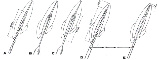
“Step back” technique. This technique is offered for treating curved canals. The aim of canal treatment is conical preparation of the root canal with the use of reciprocating motions and larger files than those used in common techniques. This technique that includes further antiseptic treatment enables to obtain clean root canals. However, it is hard to master this technique and it has the similar disadvantages to those of the coronal-apical method (possible intrusion of the used material into pre-apical tissue, block of the root apex and deviation of the initial length, especially while working with thicker and less flexible instruments).
The application of any type of endodontic instruments has its advantages as well as disadvantages. For instance, H-files are less likely to move the used material towards the root apex but if used inaccurately they may cause straightening or even perforation of the canal.
Pic. Expansion of the root canal procedure for “Step-Back” technique with increasing indentation step.
It is only the “step back” technique that enables to form the canal in the original conical shape.
Coronal-apical method
Standard technique. The coronal-apical method provides treatment and extension of the root canal from the orifice to the root apex opening, using the instruments that gradually diminish in size. First, the orifice and the middle thirds of the root canal are prepared. Then the canal working length is determined. Only after that the apical part of the canal is treated and the stop prep is made.
Coronal-apical methods are indicated in the following cases:
- in case of significant infection of the root canal content, when there is risk of pushing the pulp decay beyond the apex;
- provided machine methods of canal extending are used, e.g. if a straight canal is extended with peeso-reamers;
- if machine nickel-titanic profiles or GT-files are used.
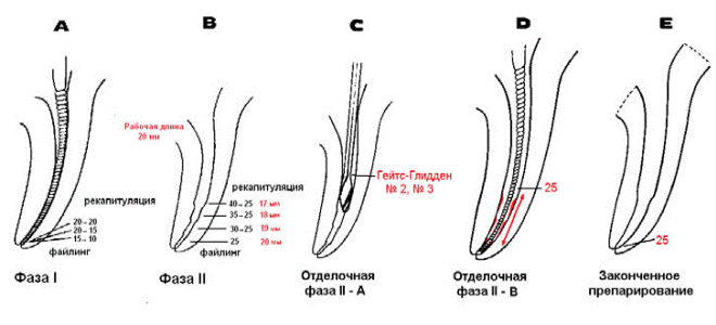
“Crown down” technique. This technique suggests phased treatment of the canal from its orifice to the apex with consistent change of instruments from bigger to smaller size (Pic.).
According to “crown down” technique mechanical treatment of the canal is carried out as follows. Firstly, the root canal penetration depth is determined with K-file № 35. If this depth is over 16mm, the coronal part is first to be prepared. In case the penetration depth is less than 16 mm, a radiograph is made. Provided the cause is in the root canal narrowing, it is necessary to prepare the canal up to the depth of 16 mm with small-sized endodontic instruments so that tool № 35 can reach 16mm. in case the narrowing is caused by the root canal curve, this canal should be prepared till the point of resistance. Next it is necessary to determine the canal temporary working length 3mm from the radiographic apex. After that K-file №35 is inserted up to the first resistance. It should be turned around twice with no apical push. This manipulation is to be repeated with a smaller tool to reach the canal temporary working length.
Рic. Expansion of the root canal procedure for “Crown down” technique.
The final working length of the canal is determined under the radiograph. Then the abovementioned stages are repeated with K-files №40 and №50 to reach the working length and the required diameter.
The canal walls are leveled with an H-file, if necessary.
“Step down” technique. This is a modified method. At the first stage the root canal coronal part is prepared with H-files № 15, 20 and 25 up to the depth of 16-18mm or up to the canal curve. Instruments № 8 and 10 are applied first of all in narrow, complex root canals. Owing to this it is possible to control the canal accessibility. H-files must be used again to double-check the accessibility of the root canal. After that “gates glidden” drills are used to shape the canal orifice. “Gates glidden” drills №3 should be inserted into the canal by 1-2mm. Next the tooth working length is determined and later, following the same procedure, the root canal apical part is prepared.
The apical opening is exposed in case it is indicated, manually or with an ultra-sound root canal hand file and the correspondent appliance.
“Crown down” or “Step down” techniques enable to smooth some drawbacks of the “step back” technique.
The advantages of coronal-apical methods are as follows:
- Provision of good access to the canal apical part;
- Reduction of the risk of infecting pre-apical tissue due to the gradual removal of decay from the canal;
- Facilitation of canal medical treatment;
- Decrease of the risk of the tool sticking in the canal apical part;
- Reduction of the risk of blocking the canal apical part by soft tissues and dentine particles;
- Decrease of the risk of “working length loss”;
- Saving the canal anatomic shape.
The drawback of this method is in the fact that at the beginning of work it is impossible to accurately determine the canal accessibility and working length.
It should be borne in mind that preparation and treatment of the canal apical part depends on the technique of the root canal obturation. If the doctor opts for the root canal filling technique with the use of gutta-percha post, then the creation of the stop prep is a crucial moment in preparing the canal. The apex of a gutta-percha post, touching the stop, must tightly obdurate the root apical opening. A bench on the root canal wall may be made using files of two or sometimes three sizes on the same depth. Thus a well-shaped bench is formed and can be used as a reliable stop for a post (fig. 7.62).
Treating very narrow root canals it is recommended to apply chemical extension. For this purpose 10-20% EDTA solution or derived gels may be used. During penetration and tool treatment of root canals chemical substances for extension must be used in 100 % cases.
Optimal time of EDTA action in the tooth canal is 15 min.
EDTA is produced in liquid form as Ededat solution (“Pierre Rolland” company), Largal ultra (“Septodont” company), and in the form of gel: Canal plus (“Septodont” company), Rc-prep (“Premier” company), File eze (“Ultradent” company), Chela-jen (“Jendent-fi” company), canal glade (“Raduga-R” company) etc. (fig. 7.64). Besides its direct dissolving action on dentine, these medicines create lubrication for instruments in the canal, which considerably reduces the risk of its sticking and breaking off.
The extension of the root canal, especially in its apical part, has minimum and maximum values. It has been found out that the minimum diameter of the canal is file №25 according to ISO. This canal extension secures more or less acceptable conditions for its filling.
In the specific clinical situation the degree of extending the root canal is set individually. Nevertheless, there are recommendations regarding the sizes of the extension of the root canal apical part to be followed (table).
Table Sizes of recommended extension of root canal apical part
| Teeth | Recommended extension of canal apical part, mm |
| Upper jaw | |
| Central incisor | 70-90 |
| Lateral incisor | 60-80 |
| Canine tooth | 50-70 |
| First premolar (two roots) | 35-70 |
| Second premolar | 60-90 |
| First molar: | |
| Frontal buccal canal | 35-60 |
| Greater palatine canal | 80-100 |
| Second molar: | |
| Frontal buccal canal | 35-60 |
| Back buccal canal | 40-60 |
| Greater palatine canal | 80-100 |
| Lower jaw | |
| Central incisor | 45-70 |
| Lateral incisor | 45-70 |
| Canine tooth | 50-70 |
| Fist premolar | 50-70 |
| Second premolar | 50-70 |
| First molar: | |
| Front canals | 35-45 |
| Back canal | 60-80 |
| Second molar: | |
| Front canals | 35-45 |
| Back canal | 60-80 |
Control questions:
1. Technique of removing pulp from the root canal treatment of pulpitis.
2. Technique of removing decay of tissue from the root canal in the treatment of periodontitis.
3. Step-Back and Crown-Down - art enlargement of root canals.
4. The sequence of endodontic instruments for root canal enlargement.
5. Tools and techniques of opening the apical hole.
6. Drugs used for chemical expansion of root canals.
7. Means to stop the bleeding from the root canals.
8. Drugs used for drying root canals.
Homework:
Independent out-of-class work
1. Prescription drugs used for treatment of canals in the treatment of pulpitis and periodontitis (antiseptics, enzymes, antibiotics, etc.).
Tests for self-monitoring and self-correction the original level of knowledge:
What form takes root canal after the expansion by «Crown-down» method?
A. the shape of the bell;
B. conical;
C. triangular;
D. rectangular.
2. Go to the apical-coronal root canal enlargement technique include:
A. «Step Down»;
B. standard technique and «Step Down»;
C. standard technique and «Crown Down»;
D. «Crown Down» and «Step Down»;
E. standard technique and «Step Back».
3. By the coronal-apical root canal enlargement technique include:
A. «Crown Down» and «Step Down»;
B. standard technique and «Step Down»;
C. standard technique and «Crown Down»;
D. «Step Down» and «Step-Back».
E. «Step-Back" and «Crown Down».
4. The first stage of the instrumental treatment of root canal:
A. Antiseptic
B. Expansion of the apical openings
C. Determination of the length of root canal
D. Expansion of the mouth of the canal
E. Disclosure of the tooth cavity.
5. Endodontic instrument size 035 has color-coded:
A. White,
B. Yellow,
C. Blue,
D. Red,
E. Green.
6. Size of instrument with a white color-coded by ISO:
A. 010
B. 015
C. 020
D. 025
E. 030
7. Tool size 025 for Standardization ISO is color-coded:
A. White
B. Yellow
C. Blue
D. Red
E. Violet
8. To extend the orifice of the root canals are used:
A. K-file
B. H-file
C. Fissure boron
D. Gates Glidden
E. Spreader
9. The last stage of the instrumental treatment of root canal:
A. Antiseptic
B. Expansion of the apical openings
C. Determination of the length of root canal
D. Expansion of the mouth of the canal
E. Disclosure of the tooth cavity
10. «Master-file» - is:
A. file, which was finished processing the mouth of the canal
B. file that is to smooth the walls of the root canal
C. file, which was completed processing of the apical part of root canal
D. file, which was finished processing the middle part of the root canal
E. file intermediate size
11. What size is an endodontic instrument with a yellow pen?
A. 015
B. 020
C. 025
D. 030
E. 035
12. What is the graphic symbol has K- file?
A. ◘
B. ●
C. ▲
D. 
E. ■
13. Instrument, which size 010 has color-coded:
A. White
B. Yellow
C. Blue
D. Red
E. Purple